
Updated April 13th, 2026
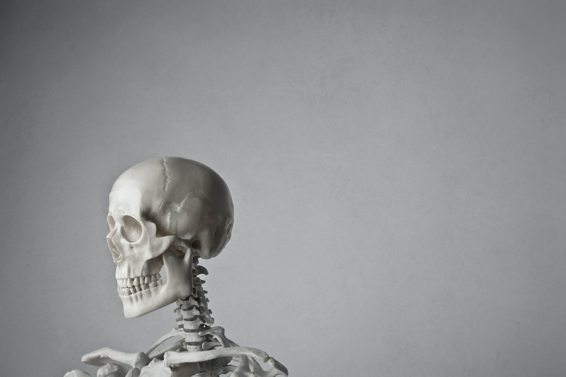
Human skeletal anatomy is a fascinating and complex construct. However, learning and fully understanding it can be a difficult process.
We all know that there are many great anatomy books out there. However, sometimes a 3D model can speed up the learning process and help you understand spatial references much better. During med school, I learned antaomy using a human skeleton model.
The knowledge, I gained still helps me every day in my clinical practice as a radiologist. In this article we will discuss important features and specific examples of the best human skeleton models to learn anatomy.
Further Reading:
Finding the Best Human Skeleton Models – The Top 5
What are the Key Features of the Best Human Skeleton Model
Depending on your needs, different models may be the best fit for you. The best skeleton model for medical studens or other medical interested persons needs to have some key features. There are several differences in quality, material, and depiction of anatomical features such as nerves, vessels and cartilage structures.
For instance, if you are using it at home, it does not need to be as durable as if you are using it to teach students. Additionally, you need to have all the structures displayed, that you want to learn or to work with. To summarize, the key features of the best human skeleton model are:
- 1. Material, workmanship and durability
- 2. Depicted anatomical structures
- 3. Flexibility and dismountability
- 4. Size
- 5. Price
In the following sections, we will shortly discuss each of these points
1. Material, Workmanship and Durability
Some anatomy departments still have a few real human skeletons. However, most modern models are made of plastic. More precisely, these are made of polyvinyl chloride (PVC), which is a durable synthetic polymer of plastic. PVC is durable, light and corrosion resistant. Additionally, it can be easily cleaned by wiping.
If you are looking for an anatomical skeleton model, make sure the quality is up to your standards. Also look at how the joints and ribs are attached and fixed to ensure that you can study without breaking things. Although this cannot be generalized, our experience is that very inexpensive models need to be moved with more care than more advanced models.
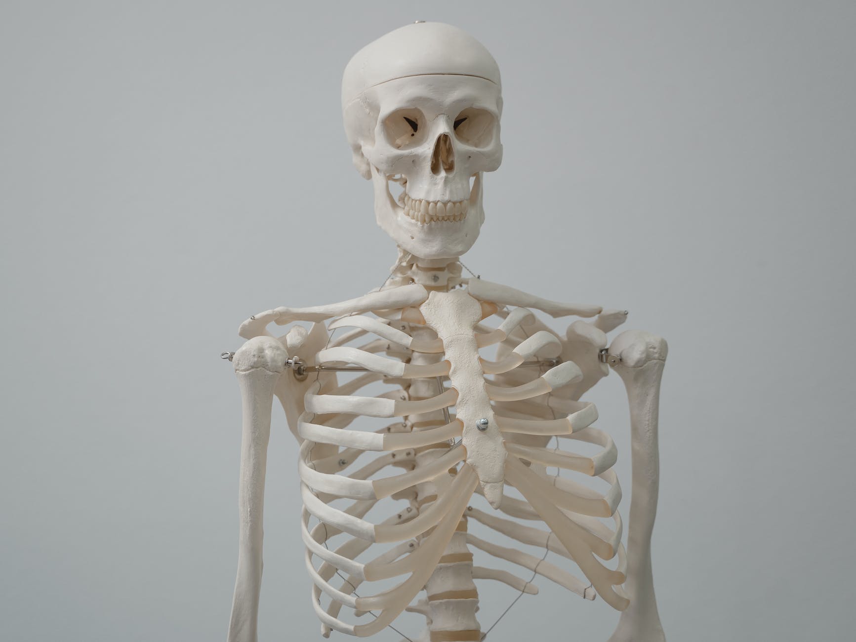
2. Depicted Anatomical Structures
Even the most basic human skeleton models come with all the major bones in the correct positions. However, there are significant differences in the additional anatomical structures represented. For instance, some models have colored muscle insertion and origin points. Others come with joint capsules and ligaments.
More advanced models even come with the whole major central and peripheral nerves and blood vessels. The best skeleton model for medical students accurately shows a lot of relevant anatomical structures. Modern high-quality detailed skeleton models are almost as good as real human skeleton for medical students in terms of detail. This may be even more important if you plan to go into anatomy-heavy disciplines such as radiology.
Be aware that more detail often means a higher price. Especially if you want a model that is anatomically correct down to the smallest detail, has correct range of motion for each joint, and shows vessels and nerves, it will most likely cost four figures.
3. Flexibility and dismountability
Because you will be touching the model and moving parts as you study anatomy, the skeleton needs to be flexible. Ideally, it will come with a good stand that also has wheels for easy movement. However, if you need to transport the model, you can also look for one that is easily disassembled and will not break when taken apart and reassembled.
Flexibility also means to be anatomically correct in funtion by accurate renderings of the human skeleton’s joints. The should all bend in the correct directions with the correct range of motion. Therefore, you will get accurate renditions of complex structures such as your hand and feet bones.
4. Size
You can get human skeleton models in a variety of sizes. For instance, there are miniature models that will fit in your pocket. However, if you really want to learn and understand the anatomical bony relations, I suggest using a life size model.
A life size anatomically correct model allows for direct comparison of complex movements between you and the model. Therefore, all models presented in this article are life size.
Think about the position of the radius and ulna when the forearm is rotated (supination and pronation). If you have a life-size model, you can move your arm and the model’s arm simultaneously. This will help you understand the parallel or overlapping positions of the forearm bones.

5. Price
As discussed in some of the points above, more details and more features usually mean a higher price. If you only need to have the basic bones with little movement, you can get one for under a houndred dollars. However, be aware that although they mostly have correct anatomical bone location, there may be downsides in movement and overall quality. If you want features such as cartilage, muscle insertions and/or nerves to be presented, a good model will range between 200 and 500 USD.
Of course, there are also high-end models used in university anatomy classes. These models have a lifelike level of detail, are durable, and may come with detailed nerves and vessels. However, these models can cost upwards of $1000 USD and are not necessarily suitable for home anatomy study.
The Best Human Skeleton Models – The Details
1. The Muscle-Man: Ultrassist Life-Size with Muscle Markers

- Good value for money
- Spinal nerves included
- Muscle insertion and origin points color-coded
- Joint ligaments depicted
- Removable calvarium, arms and legs
- Natural and realistic joint movements
Pros: Best combination of features at this price point. Muscle markers make it particularly useful for anyone who needs to understand not just bone positions but also the functional anatomy around them. The rolling stand is sturdy and well-designed.
Cons: No blood vessels. Not the most detailed depiction of small peripheral nerves.
2. The Allrounder: breesky Life-Size 70.8″

- Full life-size at 70.8 inches
- Nervous system depicted
- Comes with 3 detailed anatomy posters
- Rolling stand included
- 206 articulated bones
- Removable skull cap and fully articulated joints
Pros: The three included anatomy posters are a genuine bonus — they extend what you can learn well beyond the skeleton itself. Good overall build quality for the price, and the nervous system detail is a useful addition for anyone going into neurology or radiology.
Cons: No muscle origin or insertion markings. The joint movement on some models can feel slightly stiff initially.
3. The Detailed: NLShan Life-Size 70.8″

- 206 articulated bones cast from real human specimen
- Nervous system depicted
- Removable calvarium and fully articulated joints
- Colorful anatomy posters included
- Rolling stand with five-wheel base
Pros: Solid anatomical accuracy for the price range. The five-wheel rolling stand is particularly well-engineered and makes it easy to move between rooms. Good option if you want life-size with nervous system detail at a competitive price.
Cons: No muscle markers. Very similar feature set to the breesky model — choice between the two is largely down to personal preference and current pricing.
4. The Half-Size: breesky 33.4″

- 33.4 inches — 1:2 scale based on real human
- Metal stand included
- Removable calvarium, arms and legs
- All major joints articulated
- Every suture, foramen and groove visible
- Good level of detail for the size and price
Pros: A sensible middle ground between a full life-size model and a desktop mini. Fits on a desk or shelf without taking over the room. The metal stand is more durable than plastic alternatives at this size. Good option if you have limited space but want more than a basic miniature.
Cons: Half-size limits direct comparison with your own anatomy — one of the main advantages of life-size models. No muscle markers or nervous system detail.
5. The Desktop: breesky Mini 17″

- 17 inches — fits on any desk
- Movable arms and legs
- Removable skull cap
- Basic bone and spinal anatomy
- Very affordable entry point
- Portable — take it anywhere
Pros: The most accessible option on this list. At this price it is an easy purchase for a medical student who just wants a reference model on their desk. Surprisingly good detail for the size — the skull in particular is well-made.
Cons: No muscle markers, no nervous system, no vessels. Not suitable as a primary study tool for complex anatomy. The very basic spine and rib depiction means you will outgrow it quickly. Best used as a complement to a larger model or a textbook, not as a replacement.
Summary of the Best Human Skeleton Models for Learning Anatomy
So which one is the best skeleton model? Obviously, I cannot answer this question for everyone, as everyone has different needs. This article will give you an overview of different models with different features and price ranges. During medical school, I personally liked to have not only the bare bones but also the muscle origins and insertion points marked. Therefore, the best skeleton model for medical students shows not only the bones but also the muscles and maybe even nerves and joint structures.
However, this depends on your needs. For example, maybe you want to teach or learn different x-ray examination techniques. For this purpose, a realistic depiction of just the bones with anatomically correct movements may be sufficient. If you need to learn about multiple anatomical structures, a more detailed model may be more helpful. However you may decide, I hope this article gave you some helpful information for your informed decision.
Happy Learning!
Anatomy Everywhere – The Typical Workday in the Life of a Radiologist!
Some links on this site are affiliate links. If you choose to buy a product using these links, I might get a commision for this purchase. For you, this does not change the price.
