
Updated August 6th, 2026
Did you read kVp or kV on your radiology report? Or are you starting as a radiolographer or radiology resident? This article explains what kV and kVp in radiology stands for and what it means.
kVp = kilovoltage peak
kVp is the maximum (peak) power applied on an X-ray machine to generate X-rays .
In short
- kVp is tube voltage, the peak potential across the X-ray tube. It sets the energy of the photons, and therefore how deeply the beam penetrates and how much contrast the image has.
- Typical values run from 50 to 60 kVp for extremities to 100 to 125 kVp for an upright chest radiograph. Routine adult body CT sits at 120 kVp.
- Raise kVp by 15 percent and halve the mAs and the image density holds, the patient dose falls and the contrast flattens. That is the 15 percent rule.
- In contrast-enhanced CT the logic reverses. Lower tube voltage sits closer to the 33.2 keV k-edge of iodine, so iodine attenuates more strongly and vascular contrast rises.
- kVp controls contrast. mAs controls noise. Reaching for the wrong one is the most common exposure mistake.
Typical kVp by examination
These are the ranges you will meet on most modern equipment. Treat them as starting points, not prescriptions: the value your department uses depends on detector type, source-to-image distance, grid, patient habitus and, in CT, on whether automatic tube voltage selection is switched on.
| Examination | Typical kVp |
|---|---|
| Hand, wrist, foot | 50–60 |
| Elbow, knee, shoulder | 60–70 |
| Cervical spine, skull | 70–80 |
| Abdomen (KUB) | 70–80 |
| Pelvis, hip | 75–85 |
| Lumbar spine | 80–90 |
| Chest PA, upright with grid | 100–125 |
| Mammography (Mo/Rh target) | 26–32 |
| CT, routine adult body | 120 |
| CT, low dose chest or thin patient | 100 |
| CT angiography, paediatric CT | 70–100 |
What Is kVp or the Kilovoltage Peak? More Details please!
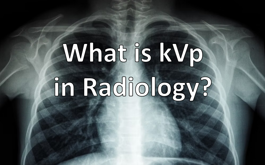
kVp Simply explained
To obtain X-ray or CT images, the X-ray machine or CT-system needs power to generate the X-rays. This power is measured in kilovolts and it determines how strong or “fast” particles (electrons) in the machine are accelerated to generate X-rays. Therefore, the kVp determines how “bright” the images will be.
The kilovolts always come in a “group” from lesser to more powerful ones. The kVp (kilovoltage peak) gives information on “how strong” the strongest parts of this power are.
What Is the Difference Between Lower and Higher kVp?
For X-ray and CT-images kVp changes how the images look:
- More kVp means stronger X-rays, more penetration and brighter images (lower contrast)
- Less kVp means weaker X-rays images, less penetration and higher contrast
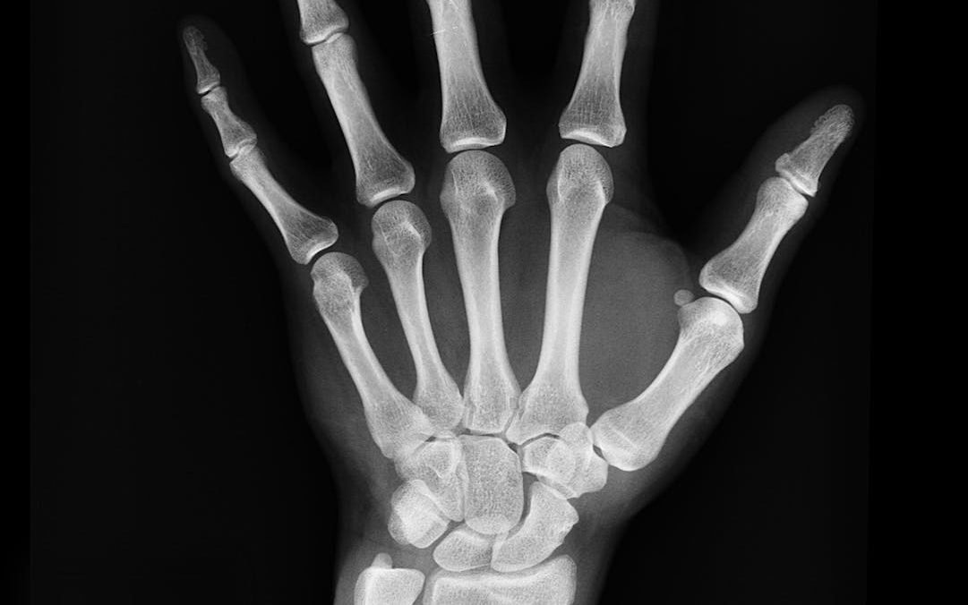
This is important for the radiologist as it determines what can be looked at in your body. For example, soft tissue (such as chest X-rays) need higher kVp (120-130 kVp). But to differentiate bones, less power is needed (70-80 kVp).
Does Higher kVp Mean More Radiation?
In brief (Answer! STAT!): If no other parameters are changed, than yes. Higher kVp means more radiation dose.
For example, in CT, the dose is almost doubled when increasing the kVp by 20% from 100 to 120 kV (if anything else is not changed).
However, there are other variables required to obtain X-ray or CT images. Therefore, the kVp cannot simply be used as a surrogate parameter for the applied radiation dose.
However, if there is higher kVp used in the room, it is even more important to have your lead glasses ready.
What Is kVp in Radiology – The More Complex Explanation
As you may know, the X-ray tube requires both a tube potential (in kilovolts, kV) and a tube current (in Milliampere-seconds, mAs). The peak potential that is applied to the X-ray tube is the kVp. This determines how fast the electrons are accelerated from the cathode to the anode. In addition, the tube voltage is responible for the amount (and quality) of the photons generated in the anode.
If the kVp is changed, the X-ray spectrum is also changed. Changing the tube voltage and kVp can significantly influence the image quality. This is especially important for anyone handeling the parameters of the X-ray or CT-machine and an important information for radiology residents. Higher kV means more penetration and therefore higher exposure (brighter image). Lower kV leads to more contrast due to lower penetration.
Further reading:
- What does STAT mean in radiology?
- What does attenuation mean in radiology?
- What does mAs mean in radiology?
The 15% rule: trading kVp against mAs
Raise the tube voltage by roughly 15 percent and halve the mAs, and the detector sees about the same exposure. The image keeps its density, the patient receives less dose, and the price you pay is contrast: higher kVp means a flatter grey scale, because the proportion of Compton scatter rises relative to photoelectric absorption.
| Starting point | Raise kVp by 15% | Then halve mAs | Net effect |
|---|---|---|---|
| 70 kVp, 20 mAs | 80 kVp | 10 mAs | Similar density, lower dose, less contrast |
| 80 kVp, 40 mAs | 92 kVp | 20 mAs | Similar density, lower dose, less contrast |
| 100 kVp, 10 mAs | 115 kVp | 5 mAs | Similar density, lower dose, less contrast |
Why CT angiography goes the other way
In contrast-enhanced CT the logic reverses. The k-edge of iodine sits at 33.2 keV, so the lower the tube voltage, the closer the mean photon energy comes to it and the more strongly iodine attenuates. Dropping from 120 to 80 or 70 kVp therefore raises vascular attenuation sharply. Noise rises too, which is why the tube current has to go up, but the contrast-to-noise ratio usually still improves and the dose falls. This is the reason paediatric and slim-patient angiography protocols run at 70 to 100 kVp while a routine portal-venous abdomen stays at 120. The dose saving from low tube voltage at preserved iodine contrast has been quantified in both paediatric and adult cohorts3.
kVp is the one parameter where the physics and the picture meet directly, and it is also where most people stop. If the way tube voltage shapes contrast interests you beyond the table above, the same logic runs through every sequence parameter in MRI, only with radiofrequency instead of photons. I built an interactive MRI course for exactly that: twenty-one units from a single hydrogen nucleus to a working scanner protocol, operated rather than read.
Frequently asked
What does kVp stand for in radiology?
kVp is kilovoltage peak, the maximum electrical potential applied across the X-ray tube during an exposure. It determines the maximum energy of the photons produced and therefore how far the beam penetrates the patient.
What is the difference between kV and kVp?
kV is the unit of voltage. kVp is the peak value of that voltage during the exposure. In practice radiographers use the two interchangeably when describing a protocol setting.
Does higher kVp mean more or less radiation dose?
Higher kVp increases dose if nothing else changes, because more photons are produced and each carries more energy. Raised together with a halved mAs, however, the net patient dose falls while image density stays roughly constant.
Why do CT angiography protocols use lower kVp?
The k-edge of iodine is at 33.2 keV. Lowering the tube voltage moves the mean photon energy closer to it, so iodine attenuates more strongly. Vascular contrast rises, and the tube current can be raised to compensate for noise while total dose still falls.
What kVp is used for a chest X-ray?
Typically 100 to 125 kVp for an upright chest radiograph with a grid. The high value is deliberate: it flattens the grey scale so that both lung and mediastinum are visible in one image.
References
- Bushberg JT, Seibert JA, Leidholdt EM Jr, Boone JM. The Essential Physics of Medical Imaging. 4th ed. Wolters Kluwer, Philadelphia, 2020.
- Bundesärztekammer. Leitlinie der Bundesärztekammer zur Qualitätssicherung in der Computertomographie. Berlin, 2023.
- Nagayama Y, Nakaura T, Oda S, et al. Radiation dose reduction at pediatric CT: use of low tube voltage and iterative reconstruction. Br J Radiol 2018;91:20170632. doi:10.1259/bjr.20170632
