
Effective dose is reported as one number in millisieverts and it is tempting to read it as the risk. It is not. Effective dose already contains tissue weighting factors, but the risk that follows from a given dose depends on who received it, and sex is one of the larger terms in that calculation.
The direction is consistent across the literature and the size of it surprises people. Here is the clearest single dataset.
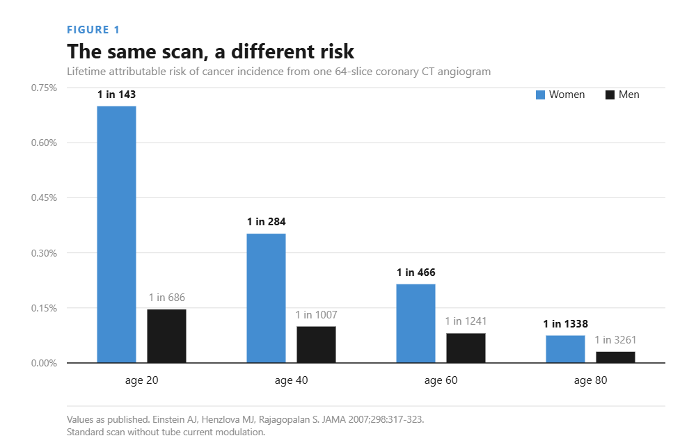
Einstein and colleagues modelled organ doses for a 64-slice coronary CT angiogram and applied the BEIR VII risk coefficients2. For a standard scan without tube current modulation, the lifetime attributable risk of cancer incidence ran from 1 in 143 for a 20-year-old woman to 1 in 3261 for an 80-year-old man1. Organ doses were 42 to 91 mSv to the lungs and 50 to 80 mSv to the female breast.
Two things in that chart deserve attention. The sex gap is roughly three to five-fold at every age. And the age gradient is steeper than the sex gradient: a 20-year-old woman carries 23 times the attributable risk of an 80-year-old man from the same examination1.
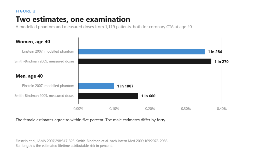
An independent estimate agrees closely. Smith-Bindman and colleagues measured actual doses in 1119 patients across four institutions and derived risks from those measurements rather than from a model phantom. They put the risk of a coronary CT angiogram at age 40 at 1 in 270 for women and 1 in 600 for men3. Einstein’s model gives 1 in 284 and 1 in 1007. The female estimates agree to within five percent; the male estimates differ by 40 percent. That spread is a fair picture of how much confidence these numbers deserve.
Where the difference comes from

Tissue in the field. Breast tissue lies inside the scan range of every chest CT, every cardiac study, every thoracoabdominal staging scan and every pulmonary embolism protocol. In women undergoing coronary CTA, lung and breast cancer together account for 80 to 85 percent of the attributable risk, and breast is the single largest contributor until age 32, after which lung overtakes it1.
Tissue weighting. ICRP Publication 103 assigns the breast a tissue weighting factor of 0.12, the same tier as lung, colon, stomach and red bone marrow4. That is not a historical constant: the 1990 recommendations had it at 0.05, and it was more than doubled on the strength of the atomic bomb survivor follow-up. The thyroid, also in or near the field of many chest examinations, carries 0.04.
Delivered dose. In one abdomen-pelvis CT series the mean effective dose was higher in women than in men, 5.76 ± 3.22 mSv against 4.37 ± 1.66 mSv5. Part of that is anatomy and part is how automatic exposure control responds to it. This is one series and should not be read as a general figure.
The variation nobody talks about
There is a fourth term, and on any given day it is larger than sex. Smith-Bindman found a mean thirteen-fold difference between the highest and lowest dose for the same study type, within and across institutions3. Median effective dose ranged from 2 mSv for a routine head CT to 31 mSv for a multiphase abdomen and pelvis.
Put plainly: which scanner your patient happens to lie on, and which protocol happens to be loaded, moves the dose more than their sex does. The sex difference is real and it is not the biggest lever available. Protocol discipline is.
What this does not mean
It does not mean withholding indicated imaging from women. A missed pulmonary embolism is a present-tense risk with a case fatality rate. A modelled cancer risk of 1 in 284 is a projection over decades. Trading the first for the second is bad medicine, and the papers that produced these numbers say so themselves.
It also does not mean the numbers are precise. Lifetime attributable risk is a model output built on the linear-no-threshold assumption, extrapolated from atomic bomb survivors exposed acutely at doses mostly above the diagnostic range. That extrapolation is contested and the uncertainty is not small: recall the 40 percent disagreement between two careful estimates of the same scan in men. These figures are useful for comparing options and close to useless as an absolute prediction for one person.
What it does mean
It shifts the threshold for a handful of decisions, and those are worth naming.
Multiphase becomes harder to justify. If a single portal-venous pass answers the question, the arterial phase must earn its place, and it earns it less easily in a 25-year-old woman than in a 70-year-old man. Note that the multiphase abdomen-pelvis protocol was the highest-dose examination in the Smith-Bindman series at a median 31 mSv3.
Alternatives get a second look. Ultrasound for suspected appendicitis in a young woman, MRI for follow-up of a known lesion, echocardiography where it answers the question. None of this is new advice. The sex difference makes it less optional.
Do not reach for a breast shield. This is the counterintuitive one. Bismuth shields degrade image quality, produce streak and beam-hardening artefact, artefactually raise CT numbers beneath the shield, and interact unpredictably with automatic exposure control, which may increase output before the shield is even in the beam. The AAPM recommends against their routine use and points to tube current modulation and iterative reconstruction as achieving comparable dose reduction with better images6. Organ-based tube current modulation, tighter collimation and a genuine reason for every phase do more than any physical shield.
Follow-up schedules compound. One scan is one scan. A five-year annual surveillance schedule is five, and if it starts at 30 rather than 60 the accumulated modelled risk is several times higher again. That arithmetic is where the sex difference becomes clinically material, not in any single acquisition.
Where it matters, and where it does not
| Setting | What it means in practice |
|---|---|
| Chest CT, young woman | Breast sits in the field and carries a high weighting factor. Justify each phase, use low kVp, skip the shield. |
| Coronary CTA | Largest sex gap in the data. Tube current modulation cuts the estimate by about a third. |
| Multiphase abdomen and pelvis | Highest dose examination measured, median 31 mSv. Ask whether one phase answers the question. |
| Surveillance imaging | Risk accumulates with each scan. Interval and start age matter more than any single acquisition. |
| Routine head CT | Breast and lung outside the field. Sex gap small: 1 in 8100 against 1 in 11,080 at age 40. |
The practical version
Dose optimisation is not a sex-specific discipline and should not become one. Every argument above is an argument for ordinary good practice: justify the study, justify each phase, use the lowest dose consistent with the diagnostic task, and prefer a non-ionising alternative where it genuinely answers the question.
What the sex difference changes is how much slack there is. For a young woman facing a long surveillance schedule, there is less. That is the whole message, and it is smaller and more useful than the headline version.
Two neighbouring questions are covered separately: why lower tube voltage lets you get away with less iodine, in the article on kVp, and why a fixed contrast volume delivers more iodine to women, in our own photon-counting data.
References
- Einstein AJ, Henzlova MJ, Rajagopalan S. Estimating risk of cancer associated with radiation exposure from 64-slice computed tomography coronary angiography. JAMA 2007;298:317–323. doi:10.1001/jama.298.3.317
- National Research Council. Health Risks from Exposure to Low Levels of Ionizing Radiation: BEIR VII Phase 2. National Academies Press, Washington DC, 2006. doi:10.17226/11340
- Smith-Bindman R, Lipson J, Marcus R, et al. Radiation dose associated with common computed tomography examinations and the associated lifetime attributable risk of cancer. Arch Intern Med 2009;169:2078–2086. doi:10.1001/archinternmed.2009.427
- ICRP. The 2007 Recommendations of the International Commission on Radiological Protection. ICRP Publication 103. Ann ICRP 2007;37(2–4). doi:10.1016/j.icrp.2007.10.003
- Estimation of radiation doses and lifetime attributable risk of radiation-induced cancer in the uterus and prostate from abdomen pelvis CT examinations. full text
- American Association of Physicists in Medicine. AAPM Position Statement on the Use of Bismuth Shielding for the Purpose of Dose Reduction in CT Scanning. full text
