
Updated March 12th, 2025
Recently, computed tomography systems with photon-counting detectors have been made clinically available. Photon-counting detector computed tomography or “Photon-Counting CT” has several technical differences to traditional CT.
In this article, we will discuss the basic principles, technical advances and clinical impact of photon-counting CT.

What Is Photon-Counting CT?
Some call it revolutionary or groundbreaking. But a photon-counting detector (PCD) is first and foremost a new detector technology for computed tomography.
If a CT system is equipped with so-called photon-counting detectors, it is called photon-counting CT.
But lets take a step back and discuss traditional and the novel photon counting detectors and their differences:
To generate an image, the CT system needs to detect the photons (or X-ray quanta) that have been generated by the X-ray tube and sent through the object (patient). This detection is performed by the CT detector.
The two detector technologies, which we would like to take a closer look at are:
- The energy-integrating detector and
- The photon-counting detector

What Is an Energy-Integrating Detector?
Traditionally, so called energy-integrating detectors (EID) have been and currently are still used in manycomputed tomography systems.
Energy-integrating detectors consist of a scintillator that converts the incident photons into light. This light is then detected by photodiodes, which convert it into an electrical signal:
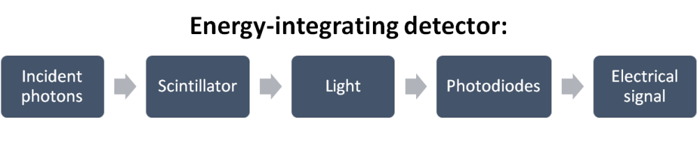
This two-step process has some limitations:
- Detector resolution: To prevent scattering of the generated light onto adjacent photodiodes, reflective septa are integrated in energy-integrating detectors. The resulting geometry significantly limits the detector resolution.
- Electronic noise: Incident X-ray quanta must have sufficient energy (>25 keV) to be detected. All photons below this threshold generate electronic noise. This noise is measurable and visible in the resulting image series
What Is an Photon-Counting Detector?
The novel photon-counting CT systems are equipped with – you guessed it – photon-counting detectors.
In terms of design, the main difference is the use of a semiconductor instead of a scintillator. In this semiconductor, the incident photons are directly converted into an electrical signal:
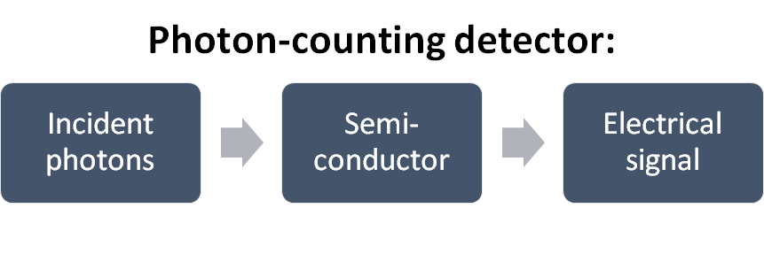
Because a strong electric field is applied to the semiconductor, this signal is then forwarded through a pixelated anode.
This process has several technical advantages over energy-integrating detectors. These advances are discussed in more detail below.
What Are the Differences Between Energy-Integrating and Photon-Counting Detectors?
Now that we talked about both CT detectors, lets put them next to eachother:

As we discussed before and as you can see in the schematic, the major difference is the material, on which the X-ray photons hit.
In energy-integrating detectors, it is a scintillater (e.g., caesium iodide, CsI). In photon-counting detectors it is a semiconductor such as cadmium telluride (CdTe) or silicone.
The photons produce light in the scintillator of the energy-integrating detector. This light scatters and has to be detected by photodiodes. In total, this is a two-step process to generate the signal.
In photon-counting detectors, the photons directly generate a signal (charge cloud) within the semiconductor. An this signal is directly conducted by the pixelated anode through a strong electrical field.
This direct-conversion process brings with it many advantages, wich we want to discuss in detail in the following sections:
What Are the Technical Advances of Photon-Counting CT?
In summary, there are 4 major advantages of photon-counting detectors over energy-integrating detectors:
- Higher resolution due to smaller detector pixels
- Elimination of electronic noise
- Equivalent detection of low-energy photons with improved image contrast
- Intrinsic spectral sensitivity with generation of inherent spectral information
1. Higher Resolution due to Smaller Detector Pixels
In classical EID-CT technology, photons are first converted into light before this in turn is converted into an electrical signal. Since the resulting light propagates spherically, a grid of reflective septa is necessary to prevent erroneous activation of adjacent detector elements (Schematic A).
On the one hand, these septa in EID-CT limit the optical resolution, since the detector pixels are limited in size due to the necessary septa. On the other hand, they also reduce the dose efficiency, since quanta absorbed by the septa do not contribute to the image signal.
In photon-counting detectors, the direct conversion of the X-ray quanta into an electrical signal eliminates the need for these separating reflective septa. Here, the incident X-ray quanta generate so-called electron-hole pairs. The electrons released as a result are diverted by the strong angel electric field and generate an electric signal on the anode pixels.
This allows significantly smaller detector pixels (currently up to 0.2 mm edge length), which result in a higher resolution.
Furthermore, the dosage efficiency is improved because there are no septa to absorb incident quanta.
Thus, the dose efficiency is only limited by the scattering grid, which is, however, indispensable for both types of detectors.
2. Elimination of Electronic Noise
In EID-CT, electronic noise is generated by X-ray quanta that do not contain the necessary minimum energy of about 20-25 keV.
This noise is inevitably included in the energy-weighted cumulative signal and is subsequently visible and measurable in the series of images reconstructed from it. The noise is amplified especially when many attenuated quanta hit the detector. This is the case, for example, with low-dose techniques or when examining patients with high BMI.
In photon-counting detectors, the specific energy of each incident photon is measured. This allows to define an exact threshold (e.g. 25 keV) below which the background noise is not taken into account and therefore completely eliminated.

The left image depicts the arrival of photons at the surface of the PCD while the right image shows a simplified version of the generated signal. Some key things to learn from this image include: the discrete nature of photon detection, the energy-dependent height of electrical pulses, and the ability to theoretically eliminate the effects of electronic noise by using a high enough threshold
Ajallphin, CC BY-SA 4.0 https://creativecommons.org/licenses/by-sa/4.0, via Wikimedia Commons, accessed 08/26/2023
By reducing or eliminating the electronic noise, the signal-to-noise ratio (SNR) is increased.
This leads to a measurably and visibly better image quality – for example in low-dose acquisitions.
3. Equivalent Detection of Low-Energy Photons
X-ray quanta with low energy are produced, among other things, by attenuation through tissue with high X-ray density (e.g. bones or iodinated contrast media).
In EID-CT, these low-energy quanta produce a relatively low light signal in the scintillator layer compared to higher-energy photons. Therefore, they are relatively devalued in the subsequent averaging process and thus have a relatively small share in the resulting signal.
In photon-counting detectors, the the energy of each incoming photon is detected.
As a result, photons with lower and higher energy photons have an energy-proportional contribution to the image signal. The allows a much broader spectrum of X-ray energy to be utilized.
In the resulting acquired image series, this is reflected in an improved image contrast.
This is particularly noticeable when iodine-containing contrast agents are used, since iodine produces an attenuation of quanta due to the high X-ray density of the contrasted vessels, especially in CT angiography. The absorption of iodine is relatively high, with a “K-edge” (K-shell binding energy for X-ray absorption) of 33.2 keV, so this advantage of photon-counting detectors is particularly prominent and results in higher iodine contrast and a higher contrast-to-noise ratio.
4. Intrinsic spectral sensitivity
In the last two decades, dual-energy computed tomography has become established for various problems in routine radiological diagnostics.
Spectral information is obtained by special acquisition techniques that allow the acquisition of multiple X-ray energy spectra of the same object. Typically, two energy spectra, one with lower energy and one with higher energy, are acquired. This spectral information can then be used to generate a series of new images in postprocessing:
Material decomposition can be performed so that, for example, iodine and calcium can be identified and quantified in the acquired data. Furthermore, virtual monoenergetic imaging (VMI) series can be calculated from these data, which give an image impression as if the object had been examined with a specific single tube voltage (monoenergetic or monochromatic). Classically, X-rays consist of a variable spectrum of energies and are thus polyenergetic or polychromatic.
Photon-counting detectors not only detect each incident photon, but also measure its energy. This results in the intrinsically available property of combining the individual energies of the quanta into so-called “energy bins” by applying certain thresholds.
For example, by applying a threshold to eliminate noise at about 20-25 keV and a second threshold at about 70 keV, the intervening energies can be combined into two energy levels. This allows all previously available dual-energy applications, such as material decomposition or the creation of virtual monoenergetic series, to be applied to each acquired scan.
Thus, no special protocols or acquisition techniques are required when using photon-counting detectors. The spectral information is inherently available for each scan due to the intrinsic spectral sensitivity.
What Are the Clinical Applications of Photon-Counting CT – Further Information?
Looking at the previously discussed four major technical advancements, lets look at how these can be utilized for clinical applications.
Disclaimer: Please be aware that this is still a very dynamic field. There are several hundred publications on photon-counting detector CT and their clinical implications (as of 09/2023). Many studies (mine/ours included) report initial experiences, phantom data or small cohorts. For many questions, initial results are available, but larger studies are necessary to confirm them. Also, this section focusses on my personal experiences and studies that I know best of.
1. Reviews of Clinical Applications of Photon-Counting CT
- Seeing More with Less: Clinical Benefits of Photon-counting Detector CT
- Technical Basics and Clinical Benefits of Photon-Counting CT
- Clinical applications of photon counting detector CT
- Photon-Counting Detector CT: Key Points Radiologists Should Know
- Exploiting the Potential of Photon-Counting CT in Abdominal Imaging
- Pediatric Applications of Photon-Counting Detector CT
- Photon-Counting Detector CT for Musculoskeletal Imaging: A Clinical Perspective
2. Clinical Applications of Inreased Resolution of Photon-Counting Detectors
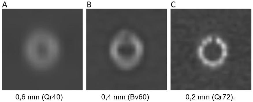
A = 0.6 mm (Qr40); B = 0.4 mm (Bv60); C = 0.2 mm (Qr72).
3. Clinical Applications of Eliminated Electronic Noise of Photon-Counting Detectors
4. Clinical Applications of Spectral Information of Photon-Counting Detectors

- Virtual coronary calcium scoring based on coronary CTA data sets.
- Improved conspicuity of liver metastases using low-keV virtual monoenergetic imaging series.
- Anemia detection and quantification from contrast-enhanced CT data sets.
For further information, also read our dedicated article on anemia detection. - Assessment of epicardial adipose tissue from coronary CTA data sets.
- Artifact reduction from dental material.
- Improved image quality of run-off CTA using low-keV VMI
When Was Photon-Counting CT first Introduced in Clinical Practice?
The short answer: Photon-Counting CT was introduced in early 2021. I had the chance to work with the third system worldwide and both with the first system in Germany and one of the first systems in the USA.
In addition, to answer this more precisely, we need to define “photon-counting detector CT” again. If you search publications on PCD-CT, you will find some before 2021. These have been performed on prototype systems. Also, some spectral systems (e.g. dual-layer CT systems) are sometimes called “photon-counting CT”. Therefore, this may lead to some confusion. However, as stated above, the first CT systems with photon-counting semiconductors in their detectors have been introduced in early 2021.
References
- Bette, S. J., Braun, F. M., Haerting, M., Decker, J. A., Luitjens, J. H., Scheurig-Muenkler, C., … & Schwarz, F. (2022). Visualization of bone details in a novel photon-counting dual-source CT scanner—comparison with energy-integrating CT. European Radiology, 32(5), 2930-2936.
- Decker, J. A., O’Doherty, J., Schoepf, U. J., Todoran, T. M., Aquino, G. J., Brandt, V., … & Emrich, T. (2023). Stent imaging on a clinical dual-source photon-counting detector CT system—impact of luminal attenuation and sharp kernels on lumen visibility. European Radiology, 33(4), 2469-2477.
- Emrich, T., Aquino, G., Schoepf, U. J., Braun, F. M., Risch, F., Bette, S. J., … & Schwarz, F. (2022). Coronary computed tomography angiography-based calcium scoring: in vitro and in vivo validation of a novel virtual noniodine reconstruction algorithm on a clinical, first-generation dual-source photon counting-detector system. Investigative Radiology, 57(8), 536-543.
- Decker, J. A., Bette, S., Lubina, N., Rippel, K., Braun, F., Risch, F., … & Schwarz, F. (2022). Low-dose CT of the abdomen: Initial experience on a novel photon-counting detector CT and comparison with energy-integrating detector CT. European Journal of Radiology, 148, 110181.
- Bette, S., Decker, J. A., Braun, F. M., Becker, J., Haerting, M., Haeckel, T., … & Schwarz, F. (2022). Optimal Conspicuity of Liver Metastases in Virtual Monochromatic Imaging Reconstructions on a Novel Photon-Counting Detector CT—Effect of keV Settings and BMI. Diagnostics, 12(5), 1231.
- Decker, J. A., Huber, A., Senel, F., Bette, S., Braun, F., Risch, F., … & Schwarz, F. (2022). Anemia detection by hemoglobin quantification on contrast-enhanced photon-counting CT data sets. Radiology, 305(3), 650-652.
- Risch, F., Schwarz, F., Braun, F., Bette, S., Becker, J., Scheurig-Muenkler, C., … & Decker, J. A. (2023). Assessment of epicardial adipose tissue on virtual non-contrast images derived from photon-counting detector coronary CTA datasets. European Radiology, 33(4), 2450-2460.
- Risch, F., Decker, J. A., Popp, D., Sinzinger, A., Braun, F., Bette, S., … & Schwarz, F. (2023). Artifact Reduction From Dental Material in Photon-Counting Detector Computed Tomography Data Sets Based on High-keV Monoenergetic Imaging and Iterative Metal Artifact Reduction Reconstructions—Can We Combine the Best of Two Worlds?. Investigative Radiology, 10-1097.
- Rippel, K., Decker, J. A., Wudy, R., Trzaska, T., Haerting, M., Kröncke, T. J., … & Scheurig-Münkler, C. (2023). Evaluation of run-off computed tomography angiography on a first-generation photon-counting detector CT scanner–Comparison with low-kVp energy-integrating CT. European Journal of Radiology, 158, 110645.
- Flohr, T., & Schmidt, B. (2023). Technical basics and clinical benefits of photon-counting CT. Investigative Radiology, 58(7), 441-450.
- Schwartz, F. R., Samei, E., & Marin, D. (2023). Exploiting the potential of photon-counting CT in abdominal imaging. Investigative Radiology, 58(7), 488-498.
- Cao, J., Bache, S., Schwartz, F. R., & Frush, D. (2023). Pediatric applications of photon-counting detector CT. American Journal of Roentgenology, 220(4), 580-589.
- McCollough, C. H., Rajendran, K., Baffour, F. I., Diehn, F. E., Ferrero, A., Glazebrook, K. N., … & Fletcher, J. G. (2023). Clinical applications of photon counting detector CT. European radiology, 1-12.
- Esquivel, A., Ferrero, A., Mileto, A., Baffour, F., Horst, K., Rajiah, P. S., … & Fletcher, J. G. (2022). Photon-counting detector CT: key points radiologists should know. Korean Journal of Radiology, 23(9), 854.
- Baffour, F. I., Glazebrook, K. N., Ferrero, A., Leng, S., McCollough, C. H., Fletcher, J. G., & Rajendran, K. (2023). Photon-counting detector CT for musculoskeletal imaging: a clinical perspective. American Journal of Roentgenology, 220(4), 551-560.
- Nehra, A. K., Rajendran, K., Baffour, F. I., Mileto, A., Rajiah, P. S., Horst, K. K., … & Fletcher, J. G. (2023). Seeing More with Less: Clinical Benefits of Photon-counting Detector CT. RadioGraphics, 43(5), e220158.
